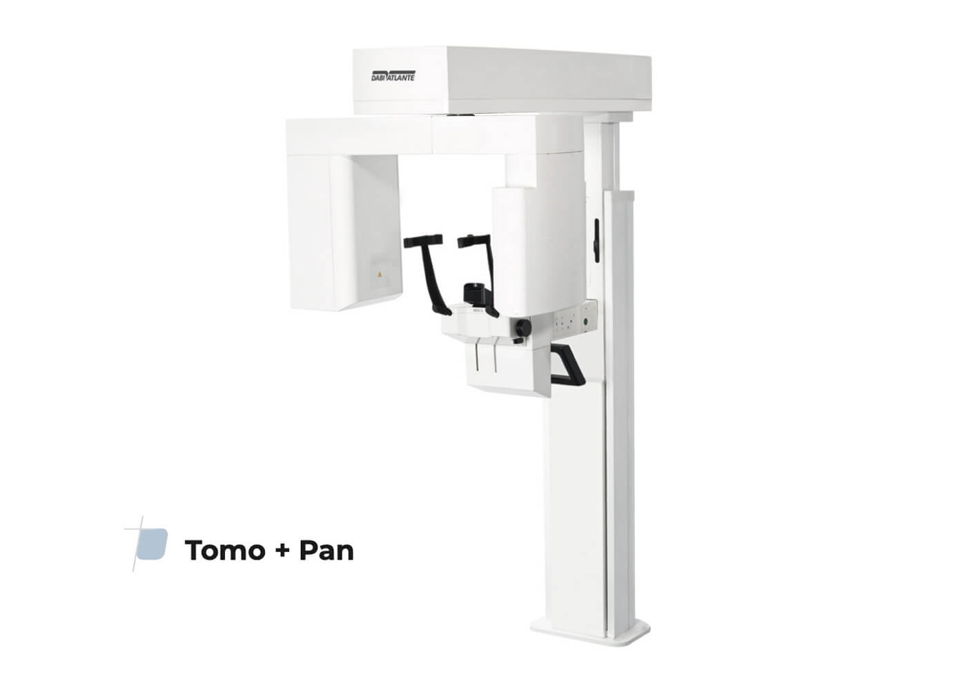
Eagle Tomo + Pan
Description
Including an exclusive Dabi Atlante’s technology and a new image processing algorithms, Dental CT Scanner AXR Eagle Edge is prepared for high patient fl ow and redefi nes innovation cutting edge, capturing images with greater precision and delivering the most accurate diagnosis, with a performance that will surprise the most experienced professionals.
Eagle Edge is designed to provide the best workflow experience to all dentists, and is designed to allow the user-friendly upgrade between its versions, offering more versatility and options. The Eagle Edge line brings all the knowledge acquired over the last 20 years, ensuring usability, hardware speed, calibration system, patient positioning, image quality and advanced image processing algorithms.
Eagle Edge is an Alliage brand of the AXR Dental Tomograph as registered by Anvisa 10101130088.
Features
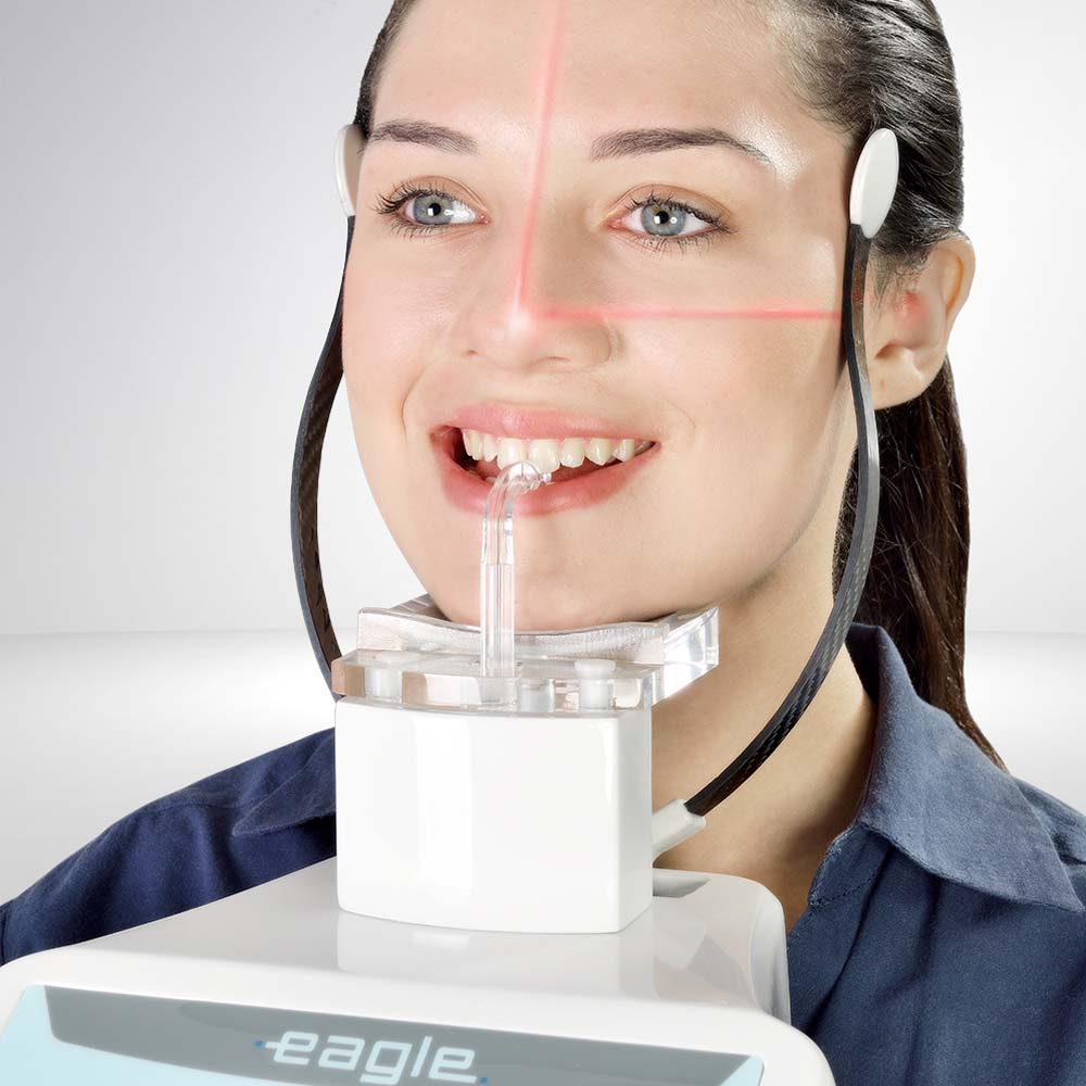
- Stability And Usability – Eagle Edge has new head positioners with 4 support points for better patient stability during exams. Simple use and easy positioning, these exclusive head positioners were designed to facilitate the clinical routine, making a faster clinic flow.
-
V-BEAM – Variable Cone-Beam – Exclusive Eagle’s Variable Cone Beam technology guarantees high definition in images with FOV of 5×5Ø, 6×9Ø and 9×9Ø as well as allowing the capture of larger images. Eagle Edge is the complete solution for 3D diagnostics, especially in applications of endodontics, implantology and orthodontics.
-
75µm Ultra High Resolution – Eagle Edge has variable resolutions with Voxel Isotropic between 75 and 400 micrometers, including automatic adjustment in relation to the size and resolution of the volume.
-
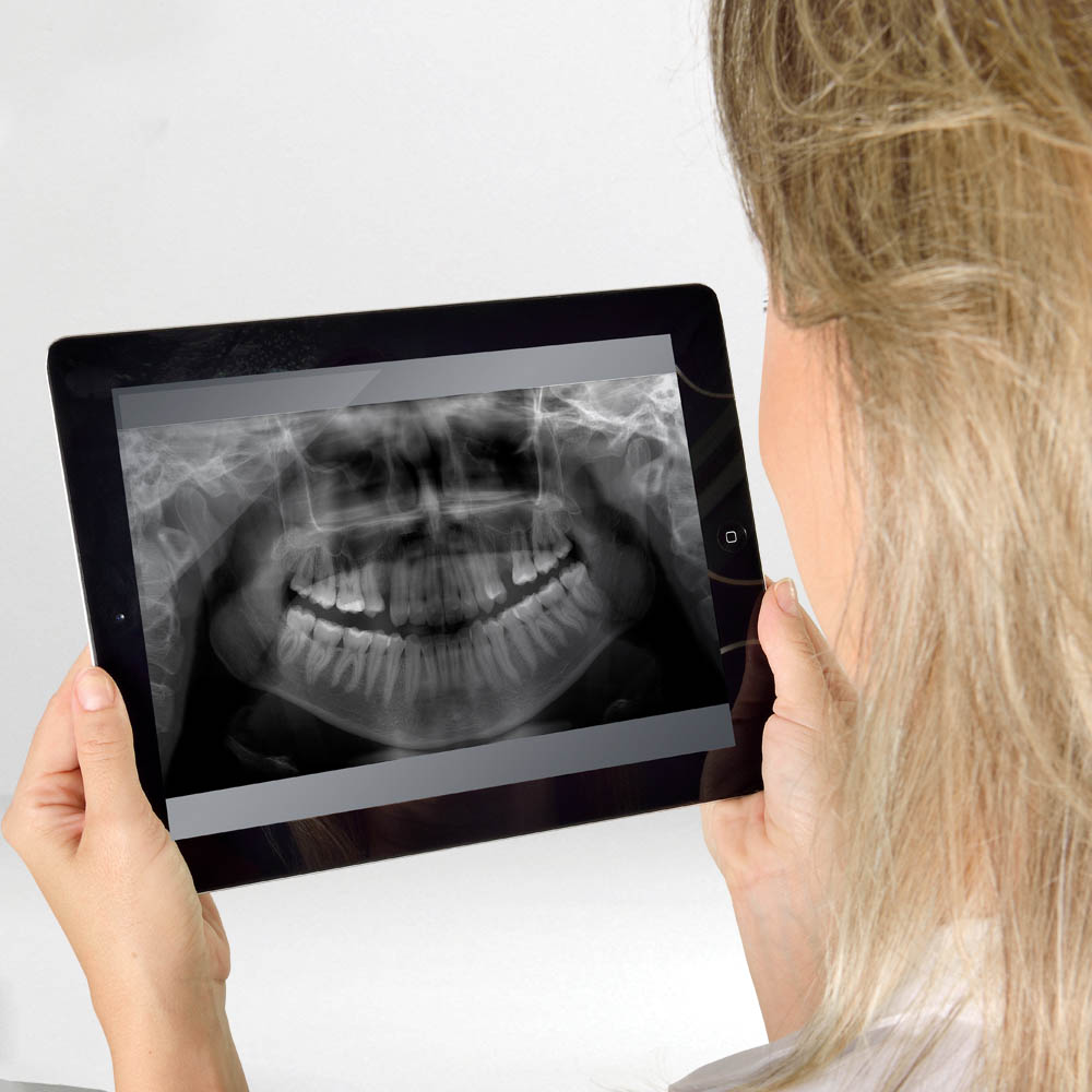
Eagle Smart Contrast – Innovative algorithm that works in all image regions, treating and improving the contrast of each area individually. The result is a homogeneous and noise-free image, allowing for details visualization and, consequently, better diagnosis.
-
Movement In All 3 Axes – The state-of-the-art movement system includes three axes(two orthogonal directions and one rotating direction) allowing greater flexibility in radiographic profiling, optimization in the cutting plane thickness and constant vertical magnification.

